The Pituitary and Pineal Glands: Structure, Function, and Modern Scientific Insights
The Pituitary and Pineal Glands: Structure, Function, and Modern Scientific Insights
The pituitary and pineal glands are small but crucial endocrine structures located deep within the human brain. Often referred to as the “master gland” and the “third eye,” respectively, their importance spans regulation of physiological homeostasis to circadian rhythms and possibly even consciousness. Here, we delve into their anatomy, function, and recent research findings that illuminate their pivotal roles.
1. Anatomy and Structure
Pituitary Gland
- Location: Housed in the sella turcica, a bony cavity at the base of the brain, beneath the hypothalamus.
- Size: ~0.5 grams, about the size of a pea.
- Lobes:
- Anterior (adenohypophysis) – glandular tissue.
- Posterior (neurohypophysis) – neural tissue.
- Blood Supply: Hypophyseal portal system, allowing direct hormonal regulation from the hypothalamus.
Pineal Gland
- Location: Near the center of the brain, between the two hemispheres, tucked in a groove where the two halves of the thalamus join.
- Size: ~5–8 mm in humans.
- Histology: Composed largely of pinealocytes and glial support cells.
- Unique Feature: Often calcifies with age (visible in radiographic imaging).
2. Functions and Hormonal Output
Pituitary Gland
The anterior pituitary synthesizes key hormones:
- ACTH (adrenocorticotropic hormone)
- TSH (thyroid-stimulating hormone)
- GH (growth hormone)
- LH & FSH (gonadotropins)
- Prolactin
- MSH (melanocyte-stimulating hormone)
The posterior pituitary stores and releases:
- Oxytocin
- Vasopressin (ADH)
These hormones influence metabolism, growth, reproduction, lactation, and water balance.
Pineal Gland
- Main Hormone: Melatonin, synthesized from serotonin in response to darkness.
- Function: Regulates circadian rhythms, influences sleep-wake cycles, and affects seasonal biological rhythms.
3. Recent Scientific Research and Discoveries
Pituitary Gland
- Stem Cell Research (2023–2024): Studies have identified SOX2+ pituitary stem cells involved in tissue repair and adaptation during physiological stress (e.g., puberty, pregnancy).
- Tumor Genomics: Advances in single-cell sequencing have uncovered distinct subtypes of pituitary adenomas, with implications for personalized treatment.
Pineal Gland
- Melatonin and Cancer (2023): Recent trials show melatonin’s antioxidant and anti-inflammatory properties may slow the growth of hormone-dependent cancers (e.g., breast and prostate).
- Calcification Studies: High-resolution MRI studies have correlated pineal gland calcification with insomnia, depression, and Alzheimer’s disease, suggesting a diagnostic biomarker potential.
4. Functional Interplay and Beyond
Emerging research hints at a neuroendocrine dialogue between the pineal and pituitary glands:
- The suprachiasmatic nucleus (SCN) receives retinal input and modulates pineal melatonin secretion.
- Melatonin, in turn, influences hypothalamic-pituitary-gonadal axis activity—affecting puberty and reproductive cycles.
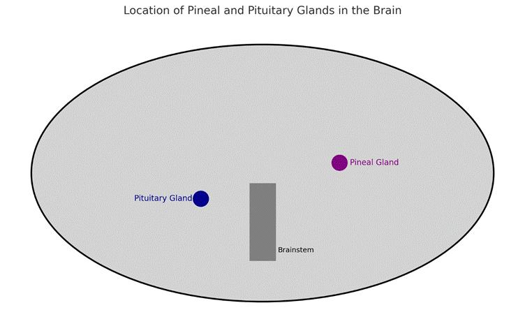
5. Visualizing the Glands
- The pituitary gland located near the base, just under the hypothalamus.
- The pineal gland situated near the brain’s midline, slightly posterior and superior.
- These are embedded in complex networks involving the thalamus, hypothalamus, and brainstem.
6. Future Directions
- Neuroplasticity & Consciousness: Some theorists and neuroscientists explore links between pineal activity and altered states of consciousness, though empirical evidence is still emerging.
- Gene Editing (CRISPR): There is potential for correcting pituitary hormone deficiencies through gene therapy.
- Artificial Melatonin Receptors: Could revolutionize sleep disorder treatment by targeting non-traditional pathways.
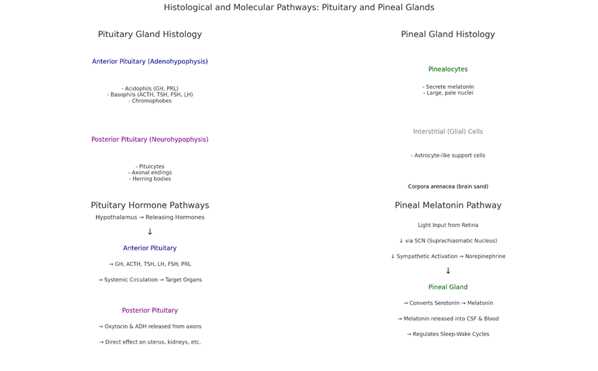
Histology
1. Pituitary Gland (Top Left)
- Anterior Pituitary (Adenohypophysis):
- Acidophils – produce Growth Hormone (GH) and Prolactin (PRL).
- Basophils – secrete ACTH, TSH, FSH, and LH.
- Chromophobes – thought to be degranulated or stem-like cells.
- Posterior Pituitary (Neurohypophysis):
- Pituicytes – glial-like supporting cells.
- Herring bodies – swellings of axons storing oxytocin and ADH.
2. Pineal Gland (Top Right)
- Pinealocytes: Main functional cells, secrete melatonin.
- Glial (Interstitial) cells: Provide structural support.
- Corpora arenacea: Calcium deposits, known as “brain sand”, increase with age.
Hormonal Pathways
3. Pituitary Hormone Pathways (Bottom Left)
- Anterior Pituitary:
- Stimulated by releasing hormones from the hypothalamus.
- Secretes hormones affecting thyroid, adrenal glands, reproductive organs, and growth.
- Posterior Pituitary:
- Direct release of oxytocin and ADH from hypothalamic axons.
4. Pineal Hormonal Pathway (Bottom Right)
- Light signals from the retina reach the pineal via the SCN.
- Darkness stimulates norepinephrine release → activates enzyme pathways in pinealocytes.
- Serotonin is converted into melatonin, which enters the CSF and bloodstream, regulating sleep-wake cycles.